Radiographs are images made by shooting x-rays through a body and onto a plate. This technique has been around since 1895, and originally used film – the x-rays will penetrate different tissues to different degrees, exposing the film to different degrees and resulting in varying shades of gray. Times have changed, though, and we no longer use film, but digital plates with sensors.
This results in much higher quality images, and these can be shared with a radiologist for a consultation. Because our radiographs are stored “in the cloud,” we send a link to your family veterinarian so that they also have access to your pet’s radiographs.

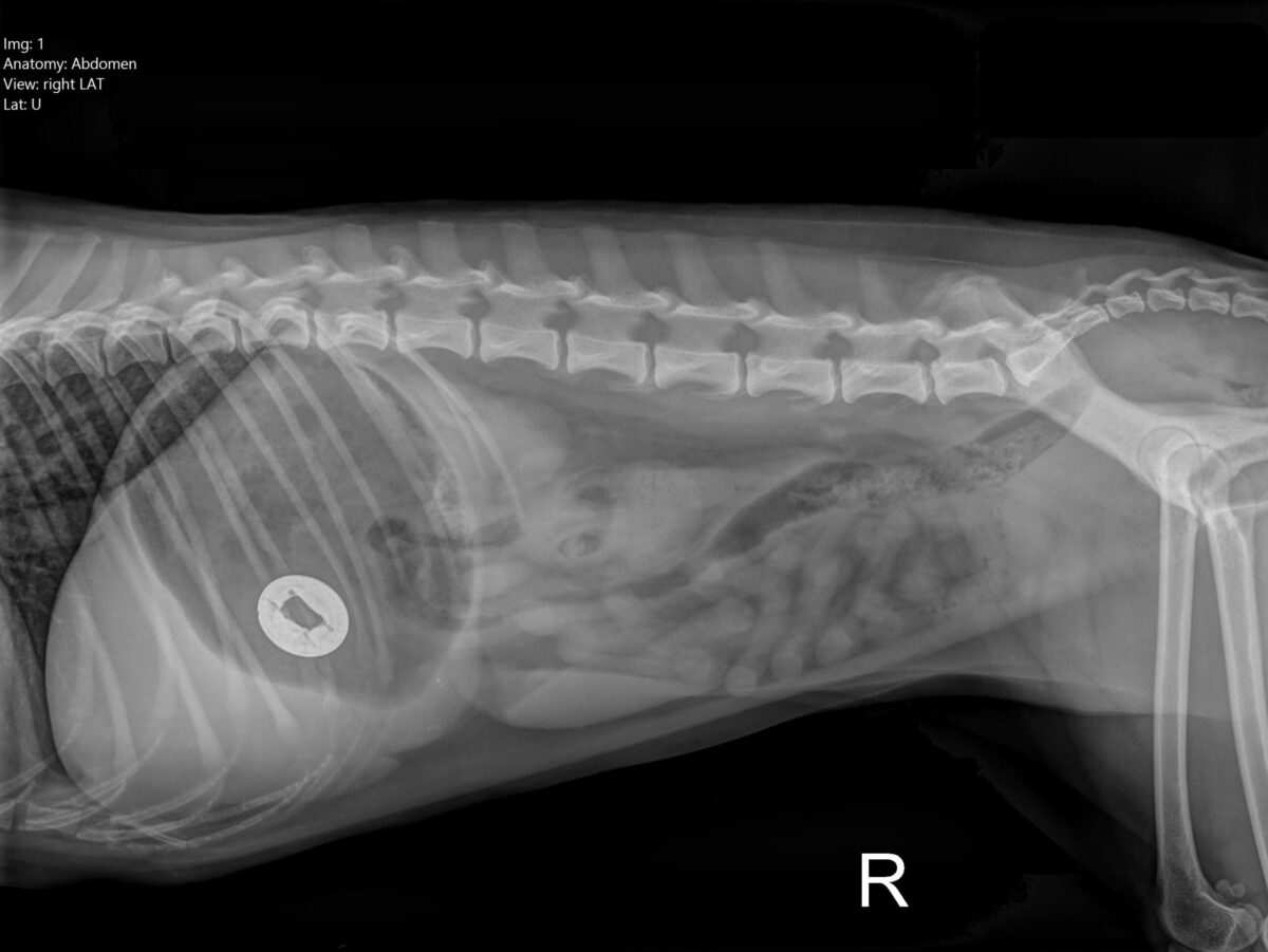
Whatever could this be in this little poodle’s stomach?
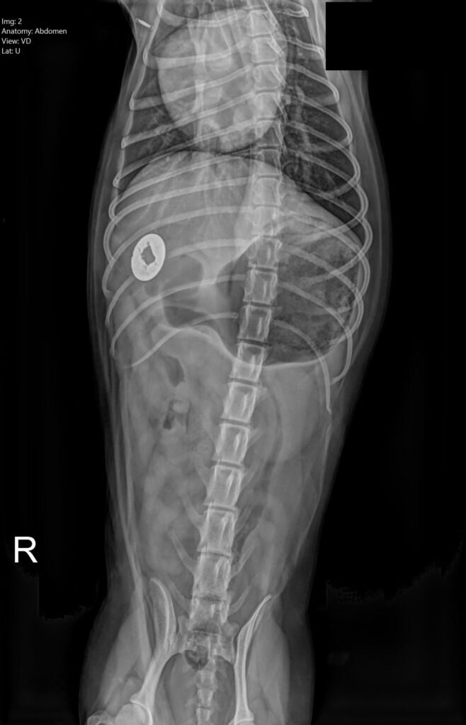
Here she is on her back, and the object can be seen where her stomach turns into small intestine.
Have you guessed yet?

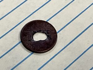
Here is the culprit – a penny! Unfortunately, the center had been drilled or punched out, which means the zinc core was exposed from the beginning (since October 1982, pennies have had a core of zinc with a thin outer shell of copper). This particular foreign body was unusual, as it was causing serious disease (zinc toxicity causes anemia, and she did need a blood transfusion) without causing an obstruction.

